
C1q and immunoglobulins mediate activity-dependent synapse loss in the adult brain
The Fc Review:
Stepping a little outside therapeutic antibody and vaccine development this week to highlight a fascinating piece of fundamental antibody biology.
A recent Science study uncovers an unexpected role for antibody-mediated complement activity in the adult mouse brain, showing that locally produced IgM and C1q work together to drive activity-dependent synapse elimination.
While this work is still early and was performed in mice, it offers an interesting look at how #antibody effector mechanisms may extend beyond the roles we typically think about.
Background:
Antibodies are typically viewed through the lens of infection, vaccination, or therapeutic development. Their effector functions, including complement activation, are well established as key mechanisms for eliminating pathogens and diseased cells.
This study asks whether antibody-mediated complement activity also contributes to normal brain physiology.
Using mouse models, the authors investigated how neuronal activity influences complement-mediated synapse remodeling and whether adaptive immune components participate in this process.
The study highlights:
- Increased neuronal activity triggered localized C1q deposition and complement-dependent synapse loss in the adult mouse hippocampus.
- Activity-dependent recruitment of antibody-secreting B-lineage cells led to local production of antigen-specific IgM, which contributed to C1q deposition at synapses.
- Reducing neuronal hyperactivity in a mouse model of Alzheimer's disease decreased C1q deposition and partially restored synaptic density.
- The findings identify a previously unrecognized interaction between adaptive immunity, complement biology, and neuronal circuit remodeling in the adult brain.
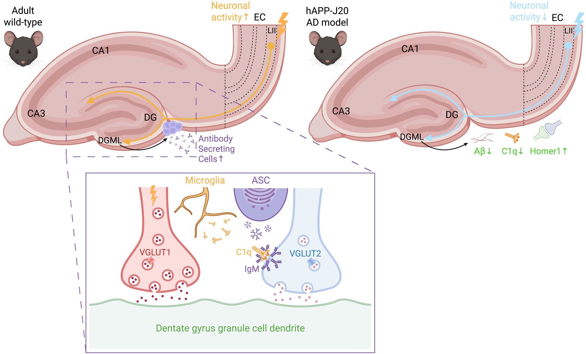
Neuronal activity modulation induces synaptic refinement through innate and adaptive immune components.
Enhancement of perforant pathway neuronal activity leads to loss of nonactivated vesicular glutamate transporter 2 (VGLUT2) presynaptic terminals. In this context, synapse loss is coordinated by release of both C1q from microglia and antigen-specific IgM from antibody-secreting B-lineage cells recruited to the hippocampus. In contrast, inhibition of perforant pathway neuronal activity induces a reduction of amyloid-β (Aβ) and C1q to restore Homer1 postsynaptic terminals. Created with BioRender.com. CA, cornu ammonis; DG, dentate gyrus; DGML, dentate gyrus molecular layer; EC, entorhinal cortex; ASC, antibody-secreting cell.
Implications for antibody development:
While these findings will need to be validated in humans, they broaden our understanding of where antibody effector mechanisms may be at work. They also suggest that adaptive immunity, complement, and microglia may work together in brain circuit remodeling in ways that were not previously appreciated.
Our perspective:
One of the reasons we enjoy this series is that it occasionally highlights discoveries outside traditional therapeutic development that may shape how we think about antibody biology in the future.
This study is a great example. It reminds us that antibody effector mechanisms continue to emerge in unexpected biological contexts, expanding our understanding of how antibodies influence health and disease. Whether those insights ultimately inform therapeutics, vaccines, or fundamental immunology, they're worth following.
References:
Gerard Crowley et al. ,C1q and immunoglobulins mediate activity-dependent synapse loss in the adult brain. Science 393, eadv1219(2026). DOI:10.1126/science.adv1219